
There are many reasons for wanting to know what goes on beneath the surface of the skin – scientists in medicine, pharmacology, and skincare use RiverD’s dedicated in vivo Confocal Raman Spectrometers to analyse the molecular composition of the skin.
Here we introduce the basics of Confocal Raman Spectroscopy, with insight into how it’s used to tell where a substance is in the skin.
Penetration problem

In our earlier posts about Confocal Raman Spectroscopy, we described how light is used to non-invasively analyse substances in the skin. This article is about the next part of the puzzle: how do we know where the substance is?
This is of particular importance to penetration studies – how deeply and quickly has a substance penetrated the skin and which layers have been reached?
When skin is illuminated with laser light, Raman scattering occurs along the entire path of the light through the skin. If all this signal were detected, spatial information would be lost; i.e. we would not know from exactly where in the skin the various Raman signal contributions arise.
Spatial selectivity
By adopting a so-called “confocal” optical configuration, Raman signal is only detected from a small tissue volume around the laser focus in the skin. In RiverD’s gen2-Skin Composition Analyzer (gen2-SCA) laser light passes through a very small pinhole and is then focused in the skin by a lens with high optical power – a high Numerical Aperture microscope objective. Raman-scattered light is collected by the same microscope objective and passes through the same pinhole on its way to a detector.
This robust confocal configuration is enabled by our near-infrared optimized microscope objective, custom-designed in-house by our team of optical designers, which achieves diffraction-limited and achromatic performance in the 650-1000 nm wavelength range.
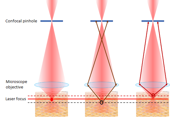
Only Raman scattered light originating from the laser focus in the skin passes through the pinhole unobstructed. Raman scattered light from above or below the laser focus in the skin is attenuated so strongly that it’s effectively blocked.

By focusing the laser light deeper and deeper below the skin surface, we can now selectively obtain Raman spectra at a range of depths in the skin. The microscope objective design has been fully optimized for skin measurements, ensuring that a 1-micron movement of the microscope objective, also results in a 1-micron displacement of the laser focus in the skin. This means we obtain depth-resolved spectra from precisely defined depths in the chosen skin layer.
Skin deep
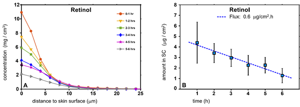
Analysis of these spectra reveals the molecular composition of the skin, visualized in profiles showing the concentration of intrinsic skin constituents or topically applied substances as a function of distance beneath the skin surface. Because these measurements are non-invasive, they can be repeated at different timepoints, e.g. to monitor diffusion dynamics of substances through the skin.
Next time we’ll look at RiverD's software tools for Raman Spectra acquisition and processing, which make measurement simple. Follow us for regular content, contact us for information about how you could use Confocal Raman Spectroscopy.