
There are many reasons for wanting to know what goes on beneath the surface of the skin – scientists in medicine, pharmacology, and skincare use RiverD’s dedicated in vivo Confocal Raman Spectrometers to analyze the molecular composition of the skin.
Here we introduce how Raman Spectroscopy identifies substances in the skin.
Unique vibrations
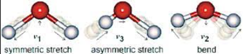
Atoms in a molecule do not have a fixed position but oscillate about their equilibrium position in so-called vibrational modes. Each molecular structure has its own specific set of vibrational modes; 3N-6 to be precise; with N being the number of atoms in the molecule.
The amount of energy needed to excite a molecular vibration is sharply determined, first and foremost by the masses of the atoms involved in the molecular vibration and the types of chemical bonds between them, but also a molecule’s secondary structure and its chemical environment play a role.
Raman scattering
When a photon; a particle of light, hits a molecule, it can transfer some of its energy to a vibrational mode. The emphasis here is on can; it is a rare event, which happens about 1 in 10 million times. When it happens, the result is that the amplitude of that molecular vibration is enhanced, and that the energy of the scattered photon has decreased by the precise amount required to excite the vibration.
This is Raman scattering, named after its discoverer, the Indian scientist Sir C.V. Raman, who observed and explained this light scattering effect in 1928, for which he was awarded the Nobel prize in Physics in 1930.
Molecular Fingerprints
Now imagine that you shine monochromatic light, consisting of photons all with the same energy, on molecules. Because of the Raman effect the scattered light will consist of a rich spectrum of lines and bands at wavelengths longer than that of the incident light.
Every unique molecular structure has its own unique Raman spectrum. This spectrum can be viewed as a molecular fingerprint; you can identify a molecule by its Raman spectrum.
A Raman spectrum of a biological tissue is comprised of the Raman spectra of all molecules present in the tissue: In fact, a Raman spectrum mirrors the overall molecular composition of a tissue.
Doubling the concentration of a particular molecule doubles the intensity of its contribution to the tissue spectrum. This proportionality is extremely useful, as it enables not only a qualitative interpretation of a tissue Raman spectrum (which molecules are present), but also a quantitative analysis (which molecules are present in what concentration).

A Raman spectrum of skin taken at a certain depth below the skin surface, not only provides information about the type of molecules present at that location, but also about their concentration. Importantly, this information is obtained, non-destructively, non-invasively and without the use of reagents. Photons only!
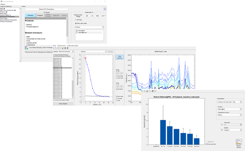
RiverD has harnessed these unique properties in its gen2-SCA dedicated Raman system for in vivo skin analysis and the SkinTools 3 data analysis software, to make light work of analyzing the skin’s molecular composition and the penetration of topical products.
Next time we’ll look at confocal selection, the technique used to provide high-resolution and to ensure concentration measurements are made at specific depths. Follow us for regular content, contact us for information about how you could use Confocal Raman Spectroscopy.