Raman Spectroscopy
Classification of cells and tissues
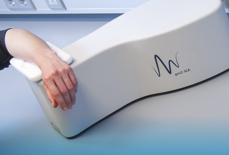
Raman Spectroscopy is a non-destructive technique, based on scattering of light by molecules. The sample under investigation is illuminated with low power laser light. In a Raman scattering event part of the energy of the incoming light is transferred to a molecule, thereby exciting one of the molecule's vibrational modes.
Because every molecule contributes to the overall Raman spectrum of a cell or a tissue, the spectrum is in fact a direct representation of the overall molecular composition. As such the Raman spectra can be used as highly specific spectroscopic fingerprints, that enable identification or classification of cells and tissues.
Data analysis
Because the technique is non-destructive, and does not require sample preparation, nor reagents, dyes, labels or other contrast enhancing agents, it is perfectly suited for in vivo application. Measurements can be carried out in a matter of seconds or less.
Raman spectra obtained with RiverD's instruments are free of instrument signatures and can therefore be obtained with high reproducibility. Data pre-processing and data analysis are carried out in real time.
Countless opportunities
In addition to its existing product portfolio, RiverD is actively developing technology and products for new diagnostic applications, based on Raman spectroscopy. We would love to hear from you and to discuss your ideas.